
Sponsored Article: Uncover More with Live Cell Imaging
Sponsored Article: Uncover More with Live Cell Imaging
Ever wondered what you might discover if you could follow the life of each cell in your live-cell assays?
Imagine if you could measure your cell’s motility without the pain and inaccuracies of having to track them by hand.
Imagine if you could also quantify how they grow, independently from how they proliferate.
Imagine if you could collect fluorescence data at the same time, all whilst minimising phototoxicity.
This is just a taste of the nirvana Livecyte offers your live-cell imaging. Phasefocus, in partnership with CELLINK, are presenting three case studies covering these different problems in live-cell imaging as part of their workshop programme at elmi2021.
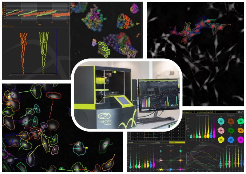
Manually tracking live-cells is a common method for deriving cell motility data. However, it is not the gold standard that many assume it to be. Small errors in click position, perhaps only a pixel or two, add up over the course of a timelapse to produce a large variability in calculated track length. Particularly when analysing full assays can take days of monotonous mouse clicking.
Livecyte can overcome these issues with its automated tracking algorithm which processes cell motility data during acquisition - meaning your cell motility metrics (amongst others!) are accurate, and ready to interpret at the end of your experiment.
An imbalance in cell growth and proliferation pathways is instrumental in several disease states including cancer and cellular hypertrophy. However, confluence and simple cell count-based assays cannot distinguish between these two different effects.
Livecyte measures a unique dry mass metric, a quantitative measure of cellular biomass, meaning cell growth can be quantified independently from cell proliferation. Additionally, dry mass can be used in the identification of mitosis events, distinguishing aberrant and asymmetric division events.
Fluorescence imaging has long played a crucial role in cell imaging, but at the same time the labelling process perturbs the cell’s natural state leaving the question of whether any observed phenotypic behaviours are due to the imaging modality.
Livecyte’s patented quantitative phase imaging (QPI) technique ptychography produces high-contrast images without the need for fluorescent labels, giving you confidence that any measured phenotypic behaviours are due to your treatments.
However fluorescence can provide some unique insights such as labelling sub-populations or to indicate protein expression. Livecyte offers correlative imaging of up to 7 different fluorescence channels. These can be captured at independent time points from the QPI modality, and the fluorescence of individual cells linked over time using the single cell tracking, enabling the long-term monitoring of fluorescence expression with substantially reduced phototoxicity.
Livecyte is a high-content live cell assay system. Join us as part of our workshop programme at elmi2021 or visit our website to find out more.