
Sponsored Article: Enhancing insights from diverse biological models
Sponsored Article: Enhancing insights from diverse biological models
Versatile microscopy systems from Olympus streamline workflows and enhance biological insights in everything from brain research to drug discovery
Innovation in imaging technology is key to scientific breakthroughs. Advanced microscopy systems and software from Olympus are designed with the needs of researchers in mind, and streamline work in a range of areas.
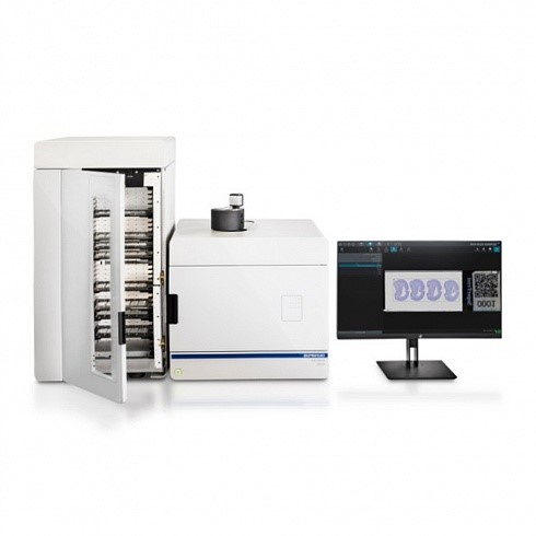
The Olympus VS200 Digital Slide Scanner
Quality for Quantification – the VS200
Obtaining sharp images from whole glass slides is essential for modern research. With features that increase image quality and throughput, the VS200 from Olympus is a fully modular, field-upgradable digital slide scanner that can accelerate brain, cancer and stem cell research and drug discovery.
By using Olympus X Line objectives with excellent chromatic correction, image flatness and high NA, the VS200 captures morphological features in high resolution – even at the periphery of an image. With oil scanning for automatic dispensing of immersion media, the VS200 also makes batch imaging easier than ever.
For optimal imaging of different sample types, the VS200 offers five observation methods: brightfield, fluorescence, darkfield, phase contrast and polarized light, which can be combined to increase the number of visible cellular structures. Fluorescence multiplexing of up to 32 channels is also possible, making the VS200 a powerful tool for studying the complex tumor microenvironment.
To increase workflow efficiency, the VS200 loader can hold up to 35 sample trays and handle slides of various dimensions. By coordinating robotic loading and unloading of trays with focusing and scanning, the VS200 also enables fast and accurate image acquisition.
To find out how the VS200 can enhance your biological insights, join our ELMI workshop on June 24th, where we explore some of the capabilities of this advanced imaging platform.
Deeper insights into 3D cellular models – the FV3000
Many of the latest breakthroughs in molecular biology are made with spheroids, organoids and tissues, which form important disease or development models. With macro-to-micro and super resolution imaging capabilities, the FLUOVIEW FV3000 Confocal Laser Scanning Microscope from Olympus helps researchers capture the complexity of 3D cellular models in more detail than ever before.
By using near infrared (NIR) lasers and silicone objectives that closely match the refractive index of living samples, the FV3000 enables precise deep-tissue imaging, giving unique insights into the internal structure of 3D specimens. NIR excitation and a high sensitivity NIR detector also reduce the required laser power – lowering phototoxicity during time-lapse imaging.
To learn more about advanced imaging capabilities of the FV3000, join our workshop on June 23rd.
For hands-on demonstrations of other advanced imaging technologies, catch our workshops on June 25th, which outline how Olympus TruAI deep learning technology is revolutionizing segmentation and classification and highlight how innovative illumination systems from Abbelight are transforming multicolor super resolution imaging.